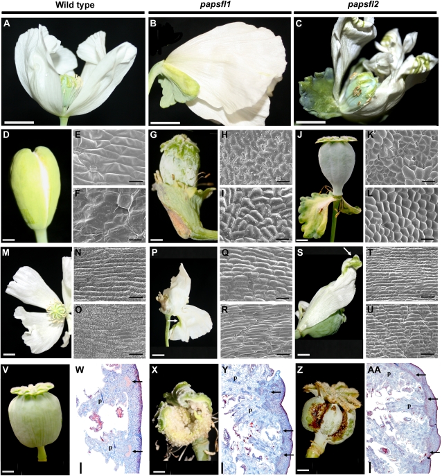
Figure 5.
Floral phenotypes in opium poppy plants treated with TRV2-PapsFL1 and TRV2-PapsFL2 separately. A, Wild-type opium poppy flower after deciduous sepals have fallen. B and C, Flowers showing persistent leaf-like sepals that remain attached to the base of the flower in papsfl1 plants (B) and papsfl2 plants (C). D, Wild-type sepals. E and F, SEM of the abaxial (E) and adaxial (F) wild-type sepal surfaces. G, Persistent papsfl1 leaf-like sepals during fruit development. H and I, SEM of the abaxial (H) and adaxial (I) leaf-like papsfl1 sepal surface. J, Persistent papsfl2 leaf-like sepals during fruit development. K and L, SEM of the abaxial (K) and adaxial (L) leaf-like papsfl2 sepal surface. M, Wild-type opium poppy petals. N and O, SEM of the abaxial (N) and adaxial (O) wild-type petal surfaces. P, papsfl1 flower showing small green patches on the abaxial surface of the outer petals. Q and R, SEM of the petaloid abaxial (Q) and adaxial (R) papsfl1 petal surfaces. S, papsfl2 flowers showing small green areas in the distal portion of the petal. T and U, SEM of the petaloid abaxial (T) and adaxial (U) papsfl2 petal surfaces. V, Wild-type immature opium poppy fruit (poricidal capsule formed by eight fused carpels). W, Cross-section of the wild-type fruit showing main vascular bundles of each placenta. X, papsfl1 fruit. Y, Cross-section of papsfl1 fruit. Z, papsfl2 fruit. AA, Cross-section of papsfl2 fruit. p, Placenta. Black arrows point to vascular traces in the pericarp, and white arrows point to green patches on the petals. Bars = 1.5 cm (A–C), 0.5 cm (D, G, J, P, V, X, and Z), 40 μm (E, F, H, I, K, L, N, O, Q, R, T, and U), 0.7 cm (M and S), and 500 μm (W, Y, and AA ).

