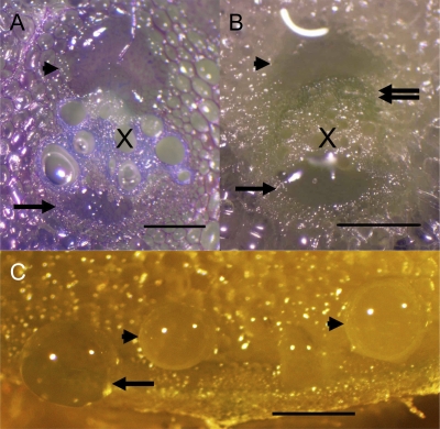
Figure 6.
Micrographs of exuding droplets. A, Cucumber vascular bundle stained with toluidine blue. The xylem (X) is stained blue, and other cells are stained purple. A small exuding droplet is seen over the external fascicular phloem (arrow). A larger droplet over the internal phloem (arrowhead) originated in the fascicular phloem but has grown to cover the extrafascicular phloem as well. B, Unstained pumpkin vascular bundle. An exuding droplet has emerged from the external fascicular phloem (arrow), which is uncommon in pumpkin. Another droplet is visible over the peripheral, internal extrafascicular phloem (arrowhead). The green region (double arrow) is the internal fascicular phloem. The white, crescent-shaped line is a diffraction image. C, Unstained pumpkin petiole section. Drops of exudate are visible over the ectocyclic phloem (arrow) and entocyclic phloem (arrowheads). Bars = 0.5 mm.

