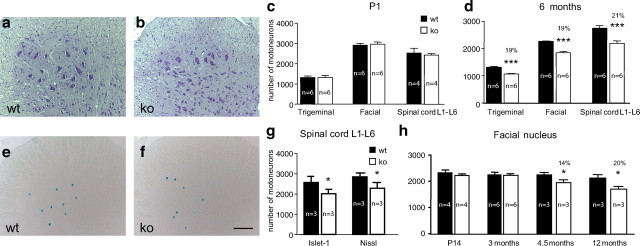
Figure 2.
Quantitative determinations of motoneuron numbers at different postnatal time points. a, b, Wild-type and knock-out spinal cord sections (anterior horn) were stained with cresyl violet. c, Counts of trigeminal, facial, and ventral spinal cord motoneurons at P1 reveal that numbers in GDF-15-deficient mice and wild-type littermates were not significantly different. d, Significant losses of trigeminal, facial, and spinal cord motoneurons in GDF-15-deficient mice, compared with wild-type littermates at 6 months of age. e, f, Wild-type and knock-out spinal cord sections (anterior horn) immunostained with Islet-1 antibodies. Scale bar, 100 μm. g, Control experiments confirm comparable losses of Nissl- and Islet-1-stained motoneurons in spinal cord sections of knock-out compared with wt mice at 6 months. h, Temporal development of losses of facial motoneurons in GDF-15-deficient mice reveals a significant decrease from 4.5 months onward and a plateau between 6 and 12 months of age.

