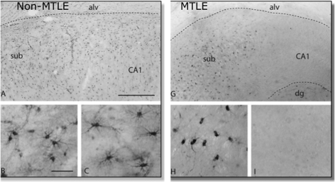
Figure 4. Expression of glutamine synthetase immunoreactivity in a non-sclerotic and sclerotic hippocampus.
Glutamine synthetase immunoreactivty in the subiculum/CA1 region in a non-sclerotic [non-MTLE (mesial temporal lobe epilepsy)] (A) and sclerotic (MTLE) (G) hippocampus. Neurologically normal autopsy hippocampus shows a pattern of staining exactly similar to (A). (B, C) in higher magnification shows GS immunopositive astrocytes in both the subiculum and CA1 area of a normal or non-sclerotic hippocampus. The sclerotic hippocampus, in which the subiculum does not have neuronal loss shows GS immunoreactive astrocytes (H), whereas the neuron-depleted astrocyte-rich CA1 area (I) shows depletion of GS in astrocytes. (A and G) Scale bar = 0.5 mm. (B, C, H and I) Scale bar = 100 μm. Reprinted from The Lancet, 363, Eid T, Thomas MJ, Spencer DD, Runden-Pran E, Lai JC, Malthankar GV, Kim JH, Danbolt NC, Ottersren OP, de Lanerolle NC, Loss of glutamine synthetase in the human epileptogenic hippocampus: possible mechanism for raised extracellular glutamate in mesial temporal lobe epilepsy, 28-37, Copyright (2004), with permission from Elsevier.

