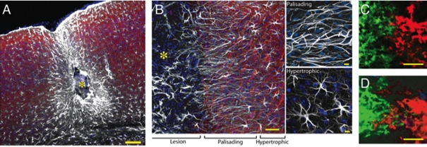
Figure 5. Organization of reactive astrocytes in a model of post-traumatic epilepsy induced by cortical injection of a ferrous chloride solution.
(A) Site of cortical injury 6 months after injury. The centre of the lesion (yellow asterisk) is surrounded by palisading astrocytes and, at a greater distance, by hypertrophic astrocytes. The mouse exhibited daily multiple generalized grand mall seizures. (B) Higher power image of a similar lesion displaying palisading and hyperthrophic astrocytes. White, GFAP; red, Map2; blue, Sytox. (C, D) Neighbouring astrocytes in control, non-EL brain exhibit little overlap of processes (C), whereas extensive overlap of processes between two adjacent astrocytes is evident in a mouse with epilepsy (D). Neighbouring astrocytes were duolistically labelled with DiL (green) or DiD (ref). Scale bar = 100 μm (A), 50 μm and 10 μm (B), 10 μm (C, D). See Oberheim et al. (2008) for details.

