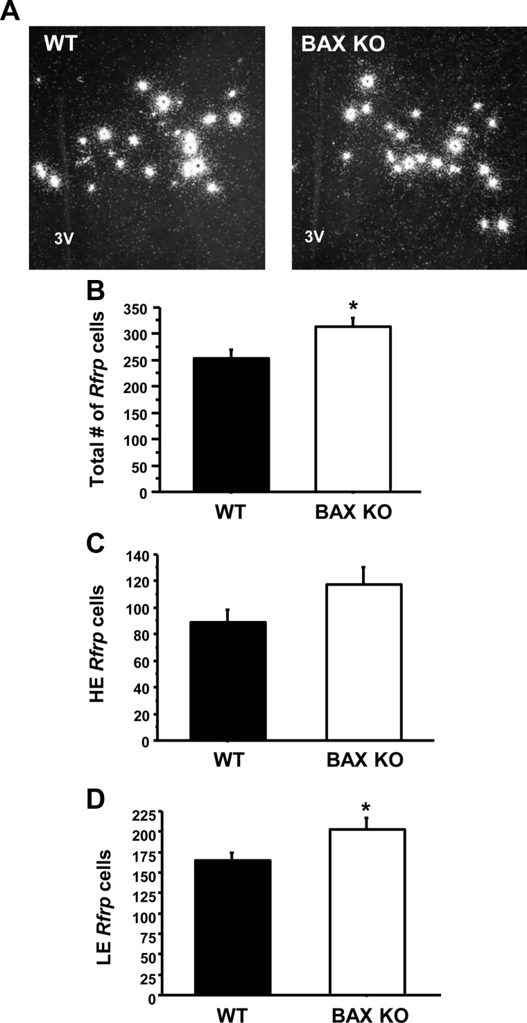
Fig. 4.
Maturation of Rfrp neurons in Bax KO mice. A, Representative photomicrographs of Rfrp expression in adult-GDX WT and Bax KO male mice. 3V, Third ventricle. B, Mean number of total Rfrp cells between WT and KO animals. *, Significantly different (P < 0.05). C, Mean number of HE Rfrp cells between WT and KO animals. There was no significant difference between genotypes. D, Mean number of LE Rfrp cells between WT and KO animals. There were significantly more LE cells in Bax KO than WT animals (P < 0.05).

