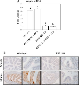
Fig. 4.
A, Expression of mRNA encoding Hpgds in whole oviducts collected from WT and ESR1KO mice. Immature mice were killed at 23 d of age (0 h) or treated with 5 IU of PMSG to induce follicular development and killed 48 h later. Data are the means ± sem for real-time PCR analysis of three samples for each treatment and genotype, with oviducts pooled from three to four mice for each sample. Levels of mRNA were obtained by real-time PCR and are expressed as fold changes. Values with different superscript letters differ (P < 0.05). B, Localization of HPGDS in the ampulla and isthmus of 10-wk-old WT, and ESR1KO mice killed on diestrus. Representative images are shown. Tissues were fixed in Bouin's fixative and embedded in paraffin. Scale bars, 50 μm.

