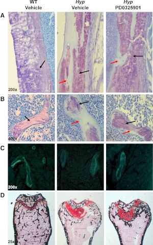
Fig. 6.
Histological analysis of the distal femur of WT and Hyp mice after treatment with vehicle or PD0325901 for 4 wk. Undecalcified sections were visualized by toluidine blue stain [purple indicates mineralized bone (black arrow); light blue indicates unmineralized osteoid (red arrow)]. A, Cortical bone section of the distal femoral metaphysis (×200 magnification). B, Trabecular bone section of the distal femoral metaphysis (×400 magnification). C, Unstained cross-sections of the femur visualized under UV illumination. Quantification of fluorochrome-labeled surfaces were performed and data were used to calculate mineral apposition rate and BFR (×200 magnification). D. Histological analysis of the growth plate at the distal femoral metaphysis in WT and Hyp mice. Plastic sections of the distal growth plate visualized by safranin O (red staining indicates cartilage) and calcified areas visualized by von Kossa (black staining) (×25 magnification).

