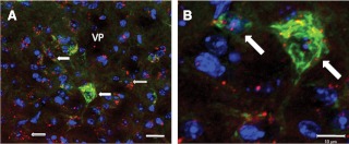
Fig. 2.
Confocal image of GH neurons in the VP. GH-IR was labeled green, and DAPI, a neuronal nucleus marker, was labeled blue, whereas DiI fluoresces red. Filled arrows indicate colocalization of GH, DAPI, and DiI, whereas open arrows indicate colocalization of DiI and DAPI only. A, GH, DAPI, and DiI colocalization in neurons within the VP (scale bar, 20 μm). B, High-magnification image of the white box in A showing a ±15 μm diameter GH neuron exhibiting DiI and DAPI colocalization (scale bar, 10 μm).

