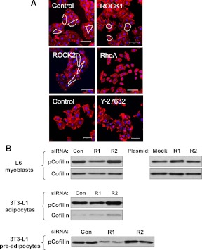
Fig. 5.
Regulation of actin cytoskeleton by ROCK1. A, The structure of F-actin in L6 myoblasts. Cells were transfected with ROCK1, ROCK2, RhoA, or luciferase (control) siRNA. Cells were treated with the ROCK chemical inhibitor Y-27632 (10 μm) for 30 min. F-actin (red) in cells was stained using Alexa Fluor 546 phalloidin. Nuclei were stained with DAPI (blue color). Representative cell morphological shapes are outlined in white. Scale bar, 100 μm (upper and middle) and 50 μm (bottom). B, Effects of ROCK isoforms on confilin-1 phosphorylation in L6 myoblasts, and 3T3-L1 adipocytes and preadipocytes. Cells were transfected with ROCK1 (R1), ROCK2 (R2), RhoA, or luciferase (Con) siRNA. Cofilin was visualized by immunoblotting with a phosphor-specific cofilin-1 Ser 3 antibody or total cofilin antibody.

