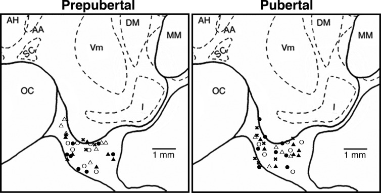
Fig. 1.
Schematic diagrams of the midsagittal plane of the basal hypothalamus showing locations of the probe tips used for microdialysis experiments in the prepubertal and pubertal stages. Most of the probe tips are located in the S-ME region. However, collection of dialysates were not limited to those spots because the microdialysis probes have a semipermeable membrane with a 5-mm length. The open and closed circles indicate morning and evening sampling experiments, respectively, in ovarian intact monkeys (experiment 1), the open and closed triangles indicate morning and evening sampling experiments, respectively, in OVX monkeys (experiment 2), and the x symbols indicate OVX monkeys treated with E2 (experiment 3). The lateral position of both prepubertal and pubertal monkeys are 0.2–1.0 mm from the midline (data not shown). AH, Anterior hypothalamic nucleus; DM, dorsal medial hypothalamic nucleus; I, infundibular (arcuate) nucleus; MM, mammillary body; OC, optic chiasm; SC, suprachiasmatic nucleus; Vm, ventromedial hypothalamic nucleus. Note that there is no significant difference in the distribution pattern of probe tips between the prepubertal and pubertal stages.

