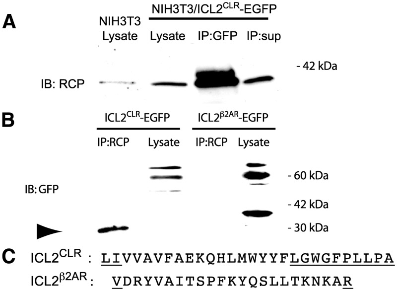
Fig. 3.
RCP coimmunoprecipitates with the second ICL of CLR. A, NIH3T3 cells expressing ICL2CLR-EGFP were lysed and immunoprecipitated with anti-GFP antibody. Immunoprecipitate (IP) was analyzed by Western blot with anti-RCP antibody. Lysate from untransfected NIH3T3 cells was loaded in first lane. For Western blots, 30 μg lysate was loaded per lane. For immunoprecipitation, 500 μg lysate was precipitated with anti-GFP antibody, and the immune complex was collected and loaded into a single lane. Supernatant from immunoprecipitation was loaded as a single lane. B, NIH3T3 cells expressing either ICL2CLR-EGFP or ICL2β2AR-EGFP were lysed and immunoprecipitated with anti-RCP antibody. The immunoprecipitate was analyzed by Western blot with anti-GFP antibody, as above. RCP monomer is indicated by arrowhead. C, Comparison of sequence of ICL2CLR and ICL2β2AR used for expression studies. Residues in putative flanking transmembrane domain are underlined. IP, Immunoprecipitation; IB, immunoblot.

