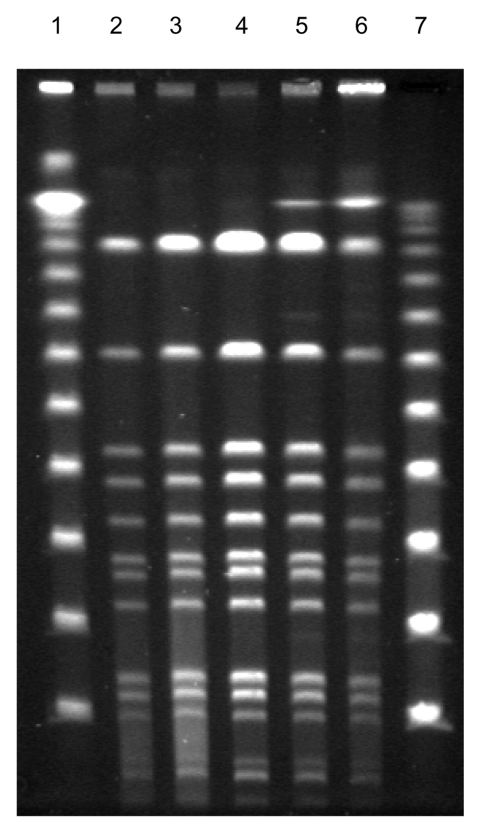
Figure 2.
Fingerprint patterns for different Staphylococcus lugdunensis colony morphotypes, including small-colony variants (SCVs), after pulsed-field gel electrophoresis after digestion with SmaI, showing identical isolates. Lanes 1 and 7, 100-bp ladder; lane 2, blood isolate; lanes 3–5, colony variants; lane 6, SCVs of S. lugdunensis obtained from thrombotic material.

