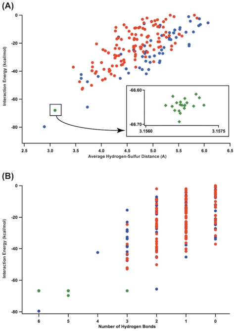
Figure 11. Hydrogen bonding environment of the 232 left- and right-handed heptapeptide-cluster conformations.
(A) Interaction energy vs. average H-S distance of left (red), right-handed (blue) complexes. Experimentally determined ferredoxin structures (green) and non-ferredoxin redox active proteins (purple) show nearly identical bond geometries and calculated interaction energies. (B). The same dataset presented as the number of hydrogen bonds versus interaction energy. Only one simulated peptide in the ensemble contributes six hydrogen bonds, corresponding to the best interaction energy. This is equivalent to the natural right-handed fold.

