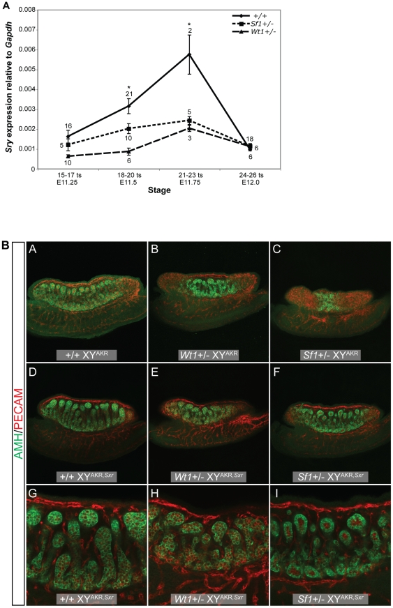
Figure 4. Sry expression is reduced in Wt1+/− and Sf1+/− B6 XYAKR gonads, but increased Sry dosage did not fully restore normal cord differentiation.
(A) Real-time qRT-PCR analysis of Sry expression relative to Gapdh (2−ΔCt) showing mean Sry transcript levels +/− standard error. The sample size for each group is shown adjacent to the corresponding data point. An asterisk denotes statistically significant effects of genotype (ANOVA, α = 0.05). There was a marginally significant effect of genotype at E11.25 (15–17 ts, ANOVA, F2,28 = 3.1670, p<0.0576) and a significant effect of genotype at E11.5 (18–20 ts, ANOVA, F2,34 = 6.999, p<0.0028) and E11.75 (21–23 ts, ANOVA, F2,7 = 15.41, p<0.0027). Pairwise comparisons revealed a significant reduction in Sry transcript levels in Wt1+/− vs. +/+ samples at E11.25 (15–17 ts, t24 = 2.224 , p<0.0179), E11.5 (18–20 ts, t25 = 3.653, p<0.0012), and in E11.75 (21–23 ts, t3 = 11.51, p<0.0014). Similarly, Sry expression was reduced in Sf1+/− vs. +/+ samples at E11.5 (18–20 ts, t29 = 2.125, p<0.0422) and E11.75 (21–23 ts, t5 = 6.142, p<0.0016). (B) The amount of tissue expressing the Sertoli cell marker AMH (green) was greater in Wt1+/− and Sf1+/− B6 XYAKR,Sxr gonads (E and F), which have two copies of the Sry gene, compared to Wt1+/− and Sf1+/− B6 XYAKR gonads (B and C), which have the normal single copy of the Sry gene (top two rows, 10× magnification with 0.7× zoom). However, while Wt1+/− and Sf1+/− B6 XYAKR,Sxr gonads developed more testicular tissue than Wt1+/− and Sf1+/− B6 XYAKR gonads, they consistently develop less testicular tissue than +/+ B6 XYAKR,Sxr controls. Additionally, cords were less differentiated and less organized in heterozygous mutants when compared to wild-type B6 XYAKR,Sxr gonads, even in the central regions of the gonads where cord differentiation was most complete (G–I, bottom row, 20× magnification). Germ and vascular endothelial cells are labeled by PECAM staining (red).

