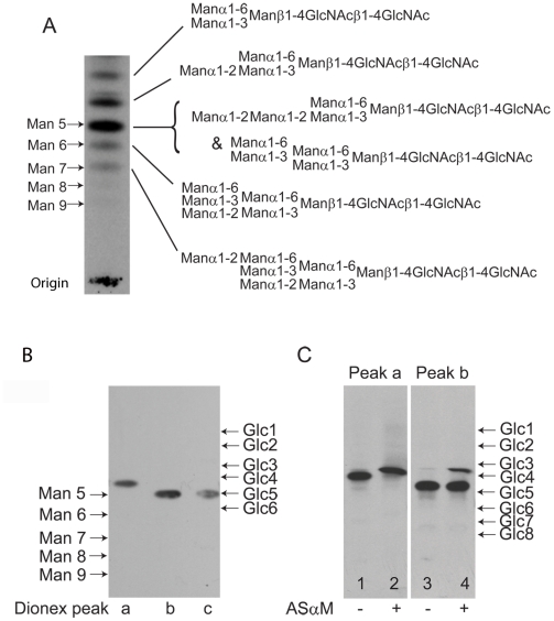
Figure 3. Fluorographs of HPTLC analyses of released and radiolabeled N-glycans from purified TfR.
(A) Total N-glycan fraction of TfR released by PNGase F and radiolabeled by reduction with NaB[3H]4. The positions of oligomannose N-linked glycan standards reduced with NaB[3H]4 are shown on the left. The proposed structures of the principal TfR glycans are shown on the right. The top three biantennary structures (Man3GlcNAc2 to Man5GlcNAc2) are of the paucimannose series and the bottom three triantennary structures (Man5GlcNAc2 to Man7GlcNAc2) are of the oligomannose series. (B) The three major components of the radiolabeled TfR N-glycan fraction isolated by Dionex HPAEC (peaks a, b and c; lanes 1, 2 and 3, respectively). The positions of NaB[3H]4 reduced oligomannose N-linked glycan and dextran oligomer standards are shown on the left and right, respectively. (C) The paucimannose Man4GlcNAc2 structure (Dionex peak a) before (lane 1) and after (lane 2) digestion with Manα1-2Man specific α-mannosidase (ASαM) and the triantennary oligomannose Man5GlcNAc2structure Dionex peak b before (lane 3) and after (lane 4) digestion with ASαM. The positions of NaB[3H]4-reduced dextran oligomers are shown on right.

