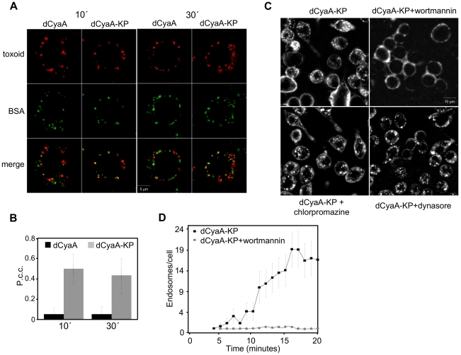
Figure 3. Inactive dCyaA-KP toxoid is endocytosed by a rapid macropinocytic pathway.
(A) J774A.1 macrophages were incubated with BSA-Dyomics 547 (50 µg/ml) together with Alexa Fluor 488-labeled dCyaA or dCyaA-KP (1 µg/ml), respectively. After 10 and 30 minutes, the cells were washed, fixed and observed as above. (B) Co-localization of toxoids with BSA was quantified using ImageJ software as described in details in Materials and Methods. (C) J774A.1 cells were incubated for 30 minutes in HBSS with wortmannin (1 µg/ml), dynasore (40 µM) or chlorpromazine (5 µg/ml), before 5 µg/ml of Alexa Fluor 488-labeled dCyaA-KP was added and time course of toxoid uptake was followed by live cell imaging and (D) quantified as above (n = 3).

