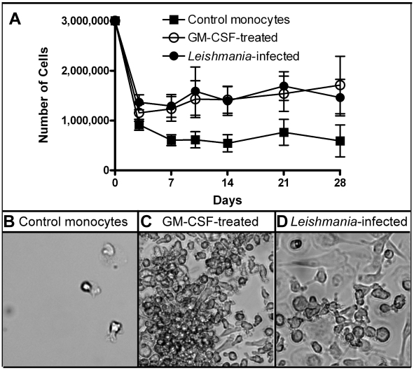
Figure 1. Determining cell viability.
A) 1×10⁶ monocytes/well were plated and then left untreated (control), GM-CSF-treated or Leishmania-infected at an MOI = 7. At various days afterwards, three wells/condition were collected, pooled and the cell numbers determined. Nine independent donors were examined. B–D) Images were captured using bright field microscope. Control cells had very few adherent cells as compared to GM-CSF-treated and Leishmania-infected cell cultures.

