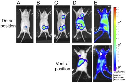
Figure 4. Different steps of bacterial spread during bubonic plague.
The bioluminescent signal was visible first at the injection site (A). It then reached the inguinal lymph node (B), the axillary lymph node (C), the liver (D, dorsal position) and the spleen (D, ventral position), and finally the entire body (E). Each picture is an example of an animal displaying a signal characteristic of each step. The color scale on the right represents the settings used to monitor light emission in all mice throughout the observation period.

