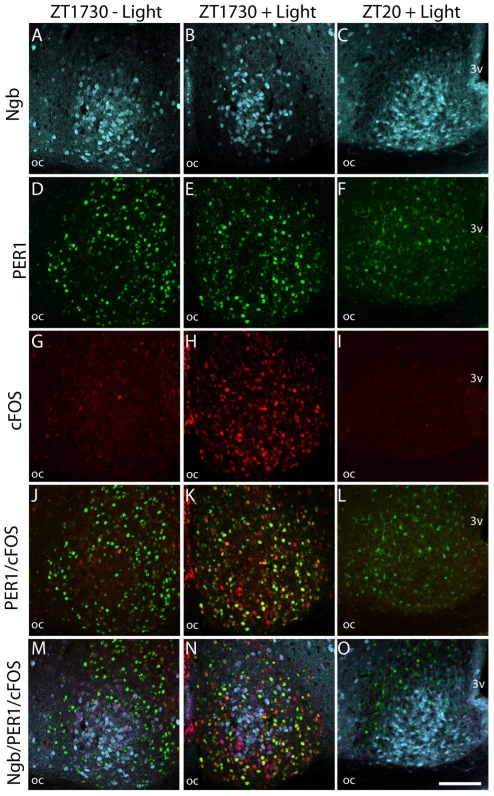
Figure 5. Light induced cFOS and PER1 in Ngb expressing neurons of the mouse SCN.
The first column shows sections of mid SCN from a mouse euthanized at ZT 1730 without light stimulation. Ngb-IR (light blue) A, PER1 (green) D and cFOS (red) G are shown. Expression of Ngb-IR and PER1, but not cFOS-IR can been observed. Merged images J–M shows PER1/cFOS and PER1/cFOS/Ngb-IR, respectively. There was no co-expression between PER1 and Ngb. Similarly, in the mid column SCN from a mouse euthanized 90 min after light stimuli is shown. Note strong expression of cFOS (H) and a high degree of co-localization (yellow) with PER1-IR (K). No co-expression could be observed between Ngb and PER1-IR, but a subpopulation of Ngb-IR cells in the core was found to be cFOS positive (N). In the last column SCN from a mouse euthanized at ZT 20 after having received a 30 min light stimulus at ZT 16 is shown. Note cFOS-IR has disappeared (I) and substantially less PER1-IR (L) is expressed in both the nuclei and cytoplasm. No co-expression between Ngb-IR and PER1-IR could be seen (O). Oc; optic chiasm; 3 V, 3rd ventricle. Scale bar 100 µm.

