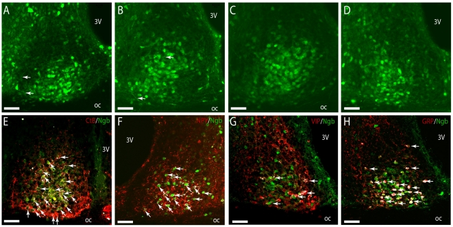
Figure 6. Innervation and co-expression of Ngb in the mouse SCN.
In A–D Ngb-IR (green) can be seen in both the neuronal cell body and processes throughout the mid and ventral part of the SCN. Highest expression of Ngb-IR was observed in the mid/core part of SCN from rostral (A), mid (B–C) and in the ventral-lateral part in the caudal SCN (D). In (E) visual input from the RHT is depicted with cholera toxin subunit B (Ctb) (red). A high degree of innervation of Ngb-IR cells (green) was observed in the ventral and mid part of the SCN shown with white colour (arrows). Likewise Ngb-IR cells were also innervated by NPY-IR fibres (red) originating from the GHT (arrows) (F). In colchicine treated mice no co-expression of Ngb-IR and VIP-IR could be seen (G). Most GRP-IR cells were seen to co-express Ngb-IR (arrows) in colchicine treated mice (H). Oc; optic chiasm; 3 V, 3rd ventricle. Scale bars 50 µm.

