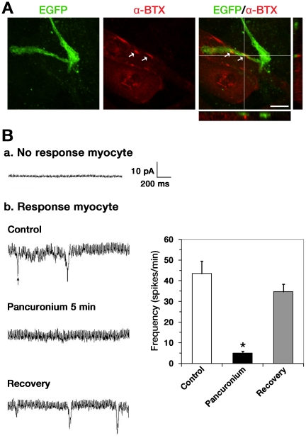
Figure 5. MNIM-EOH cells can trigger AChR clustering and form functional connections with cocultured myotubes.
(A) Fluorescence images of MNIM-EOH cells at 1 day after cocultured with C2C12 myotubes. α-BTX staining revealed AChR clustering on C2C12 muscle fibers. Confocal Z-stack imaging, in a line through the region of apparent colocalization, confirmed EGFP+ axons in close proximity to AChRs (arrows). Scale bars: 10 µm. (B) End-plate currents (EPCs) were recorded from myotubes located in close proximity to MNIM-EOH cells. EPCs were blocked from the same cell after application of 15 µM pancuronium. However, after washing out of pancuronium, EPCs could be recorded again. The bars represent mean±SEM. * P<0.05. The significance was determined by Student’s t test.

