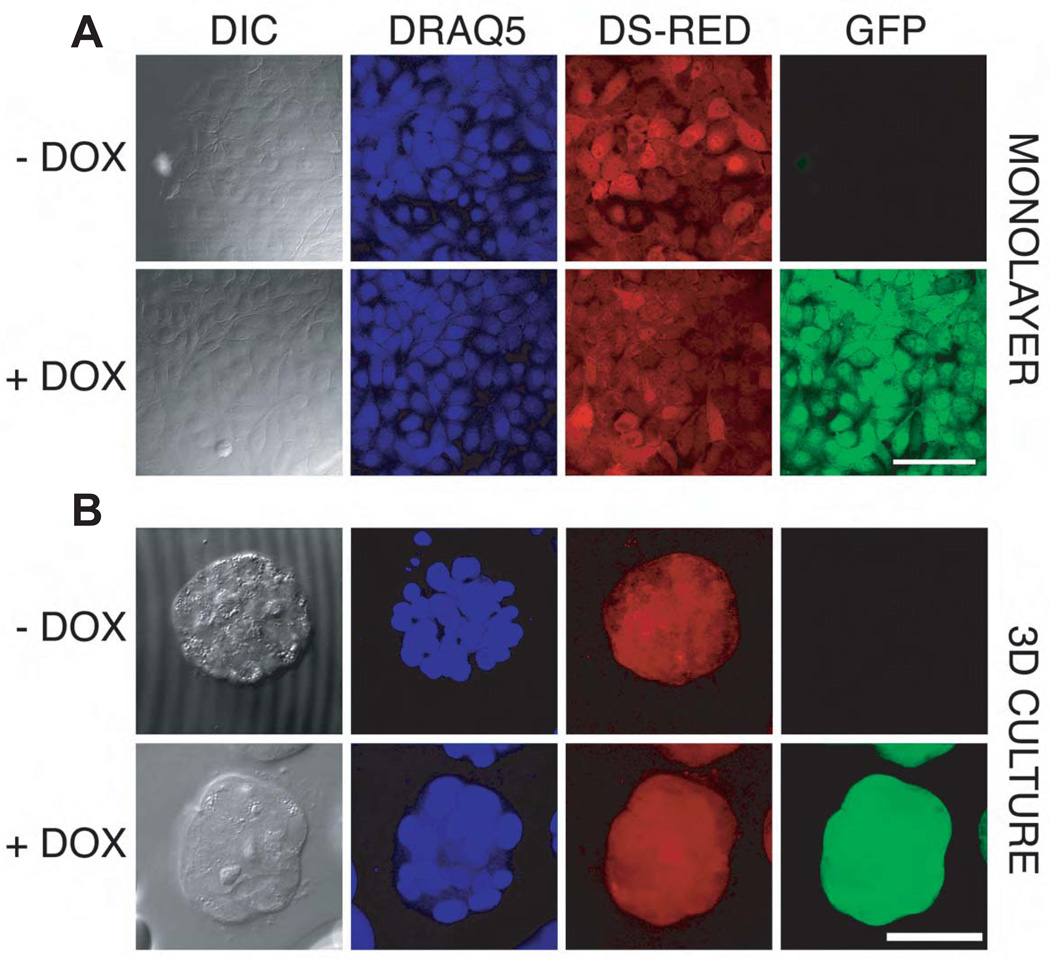
Figure 1. Doxycycline-Inducible shRNA expression in MCF-10A cells.
In this system dsRED and the TET-KRAB regulator are constitutively expressed from the same vector while GFP fluorescence is expressed only after doxycycline induction of the shRNA targeting BRG1.
Cells were sorted after 2 days of doxycycline induction (0.1 µg/mL). Then, after 5 days of culture without doxycycline, cells were seeded on coverslips and induced or not with 0.05 µg/mL doxycycline for 3 days before fixing with formaldehyde and staining nuclei with DRAQ5. Confocal image stacks, shown here for the MCF-10A-SCRAM control cell line), were collected for both monolayer (Panel 1A, scale bar = 100 µm.) and 3D culture (Panel 1B, scale bar= 50 µm.). The micrographs of the acinus (Panel B) were maximum intensity projections. Confocal settings were held constant so that linear quantitative comparisons could be made between samples with and without doxycycline induction.

