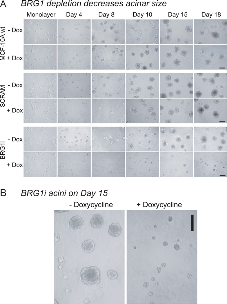
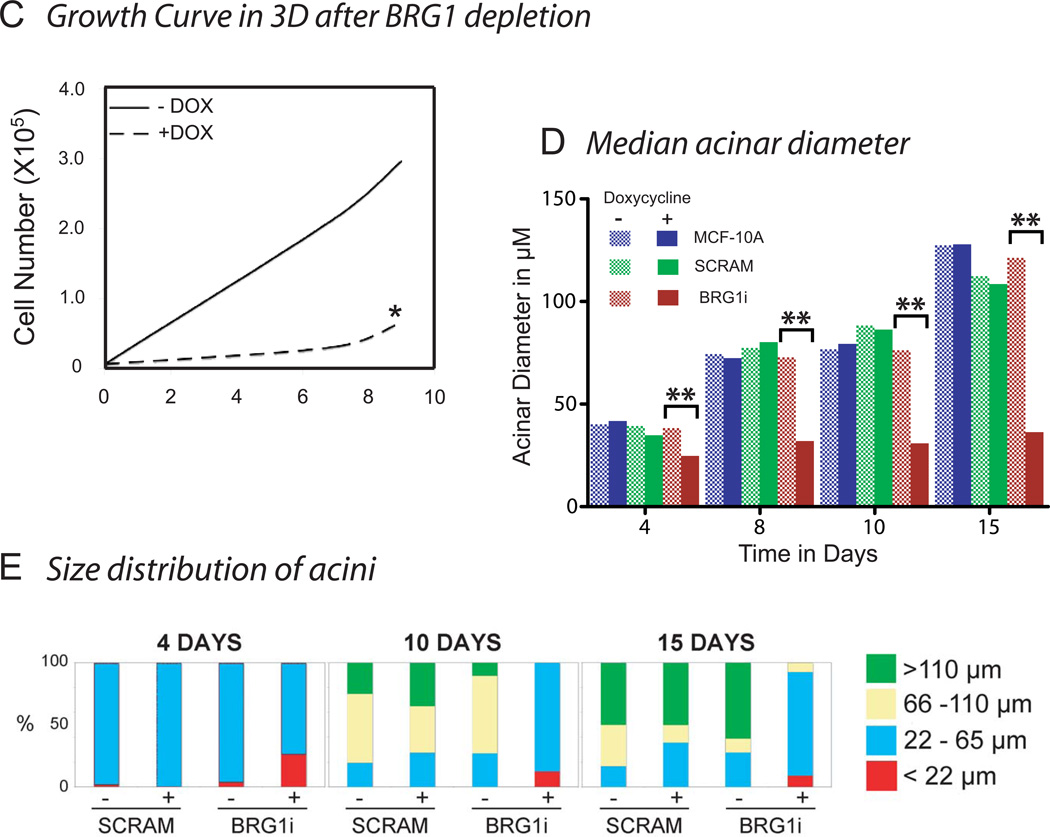
Figure 3. Decreased Proliferation after BRG1 knockdown in rBM culture.
(A) MCF-10A cells were seeded with or without doxycycline in Matrigel. The micrograph for day 0 shows the initial monolayer culture. Every two days, media with or without doxycycline were replaced and phase contrast micrographs were taken. Decreased proliferation was observed from day 4 in MCF-10A cells expressing the shRNA targeting BRG1 (+doxycycline). Size bar : 150 mm
(B) Representative micrographs of MCF-10A BRG1i acini grown in the presence or absence of doxycycline for 15 days. These pictures were among those used to make the measurements of acinar diameter presented in panel D. Size bar : 150 µm.
(C) The three dimensional Matrigel cultures were trypisinized to obtain a single cell suspension at different times after seeding (day 4 and day 9). Then, cells were counted both using a hemacytometer with trypan blue to detect dead cells, and with an automatic cell counter. The star * indicates that no increase of trypan blue positive dead cells was detected in the BRG1i cells grown with doxycycline.
(D) Median acinar diameter during differentiation. For each cell sample the median acinus size was calculated (n ≥10 acini with 2 measurements of diameter for each acinus). Statistical analysis was by a Student’s t-test. The comparisons marked ** had a high degree of statistical significance (p ≤ 0.01).
(E) Size distribution of acini. The percentage of acini with a diameter smaller than 22 µm, contained in the interval 22 to 65 µm, 65 to 110 µm, or greater than 110 µm were calculated.


