
Figure 4. A model for the spatial regulation of RhoC activity during invadopodium protrusion.
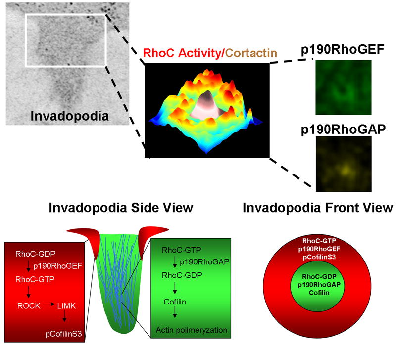
Formation of a focused invadopodium is mediated by the spatiotemporal localization of RhoC activity outside invadopodia. RhoC activity increases around the invadopodial core structure, as shown by this plot of the maximum projection over time of RhoC activity. Pseudo-color shows low RhoC activity levels (blue) to high RhoC activity levels (red) in relation to low (white) and high (brown) cortactin intensity where cortactin marks the central core of the invadopodium. This activation pattern is achieved by activation of p190RhoGEF (green) outside and p190RhoGAP inside (yellow), restricting RhoC activity just outside the invadopodium core. This spatial restriction localizes active cofilin to the core of the structure and focuses actin polymerization so as to achieve optimum protrusion elongation and invasion (bottom part adapted from [68]).