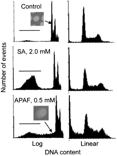
Figure 3.
Flow cytometry of rapidly proliferating mIMCD3 cells treated with SA or APAF for 4 days. Both drugs increase the percentage of particles in a subdiploid peak (under the bars), which are apoptotic bodies. In addition, APAF compared with SA or control results in the subdiploid DNA region having a greater number of cells with DNA content closer to, but still below, the G1 peak. The nuclei in those cells are intact and are swollen compared with controls (Insets, photographed in a separate experiment analyzed by LSC and with a concentration of 2.0 mM APAF).

