Figure 8.
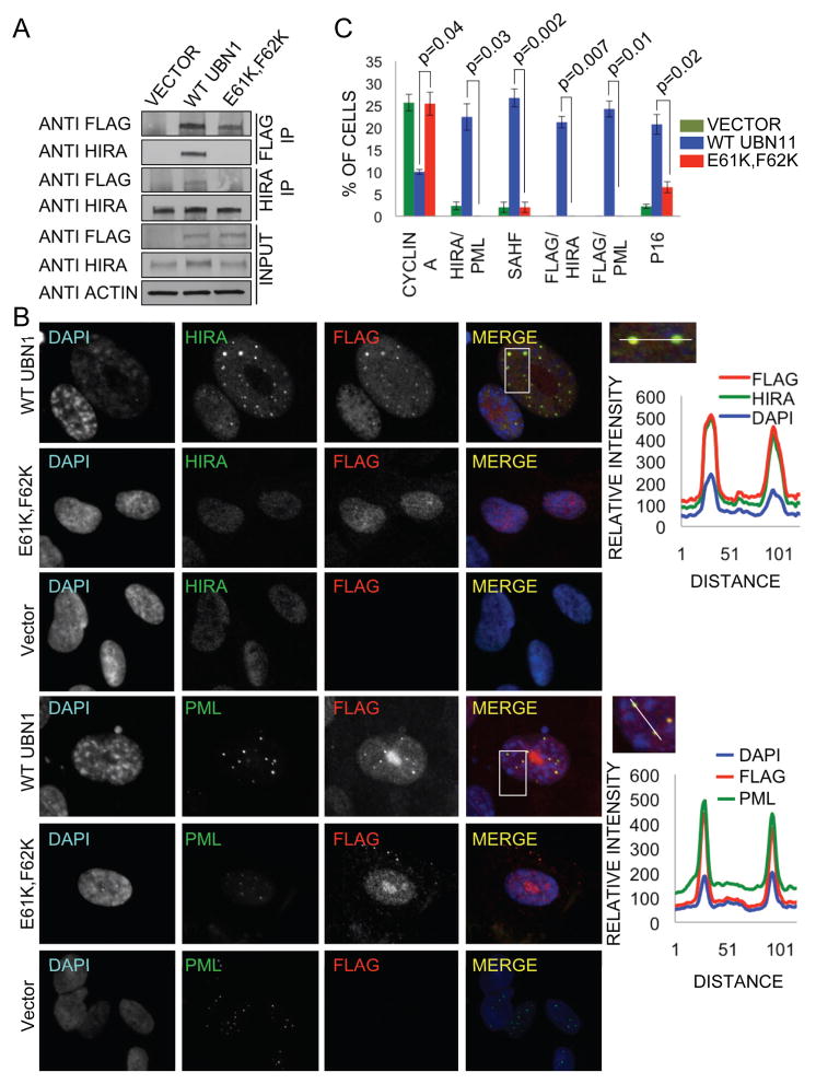
Ectopically expressed UBN1 WT, but not the (E61K, F62K) NHRD mutant, interacts with endogenous HIRA. Proliferating IMR90 cells were infected with retroviruses encoding wild-type flag-UBN1, flag-UBN1 (E61K, F62K) mutant or empty vector, as indicated. A) 5 days later, lysates were prepared and physical interactions between ectopically expressed wild type or mutant UBN1 and endogenous HIRA detected by immunoprecipitation-western blot analysis. B) 28 days later, cells were stained with DAPI and anti-HIRA (mouse monoclonal IgG1 WC cocktail) or anti-PML (Santa Cruz sc-966, mouse monoclonal IgG1) and anti-flag (Sigma F7425, rabbit polyclonal) antibodies and visualized by immunofluorescence. Upper panel on the right represents relative intensity of DAPI, HIRA, and Flag fluorescence along a straight line through the HIRA/Flag foci. Bottom panel on the right represents relative intensity of DAPI, PML body, and Flag fluorescence along a straight line through the PML/Flag foci. C) Quantitation of results from B and Figure S5. More than 100 cells were scored from 3 independent biological replicates. p values were calculated using two-tailed t-test with Bonferroni correction.

