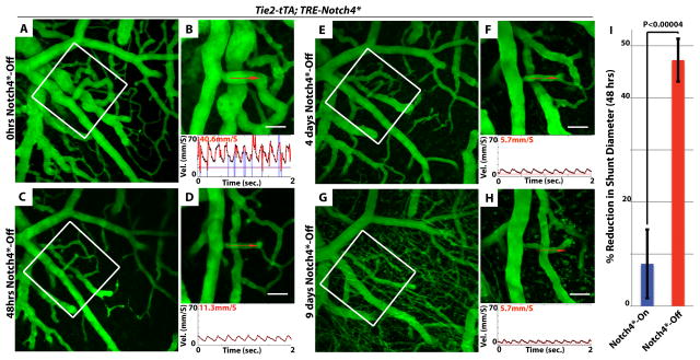
Figure 1. Repression of Notch4* induces the normalization of arteriovenous malformation.
(A–H) Two-photon timelapse imaging of cortical brain vessels through a cranial window in Notch4* mutant mice. Vessel topology was visualized by intravenous FITC-dextran. Arteriovenous shunt (A&B) was reduced in diameter following the repression of Notch4* (C–H). Centerline velocity in the regressing AV shunt was obtained by direct measurement of the velocity of individual red blood cells (panels B,D,F,H). Repression of Notch4* decreased blood flow velocity in shunt by 48hrs (compare B to D). (I) Quantification of the changes in shunt diameter without repression of Notch4* (Notch4*-On) or with repression of Notch4* (Notch4*-Off) for 48hrs. Diameter was measured at the narrowest point between artery and vein in Notch4*-On mice before and after treatment (n=22 AV shunts in 10 mice with and n =35 AV shunts in 11 mice without Notch4* repression). Error bars represent s.e.m. between individual AV shunts. Scale bars = 50μm.

