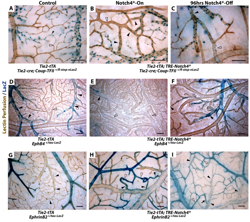
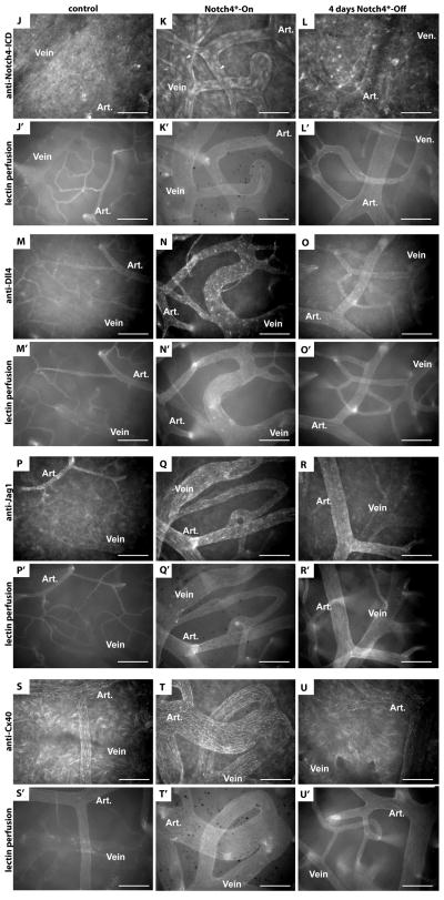
Figure 4. Turning off Notch4* normalizes arteriovenous specification in AV shunts.
(A–I) Whole mount LacZ staining of the surface vasculature of the cerebral cortex to reveal expression of Notch upstream venous specification gene Coup-TFII, downstream venous marker EphB4, and downstream arterial marker ephrin-B2. Perfused vessels were counterstained by colorimetric 3,3′-diaminobenzidine (DAB) reaction with horseradish peroxidase (HRP) bound tomato-lectin. (A–C) LacZ staining of Tie2-cre activated Coup-TFII reporter. (A) In control mice, Coup-TFII was expressed in the veins, venules and capillaries up to the arterioles. (B) In Notch4* expressing mutants, Coup-TFII was expressed in the vein, and venous portion of the AV shunt. (C) After repression of Notch4*, the narrowest point in AV shunts was found between Coup-TFII positive and Coup-TFII negative endothelium. (D–F) LacZ staining of EphB4 reporter. (D) In control mice, EphB4 was expressed in the veins and venules up to the capillaries. (E) In Notch4* expressing mutants, EphB4 expression was reduced through AV shunts, venules and veins. (F) Following the repression of Notch4*, EphB4 expression was increased in the regressing AV shunt. (G) In control mice, ephrin-B2 expression was detected in the arteries and arterioles up to the capillaries. (H) In Notch4* expressing mutants, ephrin-B2 expression was detected in arteries, the AV shunts, and at lower levels extending into the veins. (I) After repression of Notch4*, ephrin-B2 expression was decreased in the regressing AV shunts and veins. Closed arrowheads indicate venules; open arrowheads indicate arterioles. n=3 (A–C,G,I), n=4 (H), and n=8 (D–F) for each condition. Scale bars = 100μm.
(J–U) Whole mount immuno-fluorescence staining of cerebral cortex after FITC-lectin perfusion. (J–L) Endothelial localization of Notch4-ICD was undetectable in control mice (J), but present in a focal manner consistent with nuclear localization throughout the artery and vein in Notch4*-On mice (arrowheads in K), and reduced once Notch4* was turned off (L). Arterial markers Dll4 (M–O), Jag1 (P–R) and Cx40 (S–U) were expressed in the artery and not vein in control mice (M,P,S). All of these markers were upregulated in the artery, through the AV shunt, and into the vein in Notch4*-On mice (N,Q,T). When Notch4* was turned off the expression in the AV shunt and vein was lost, but arterial expression remained (O,R,U). n=5 for all mutants, n=2 for each controls. Scale bars = 100μm.


