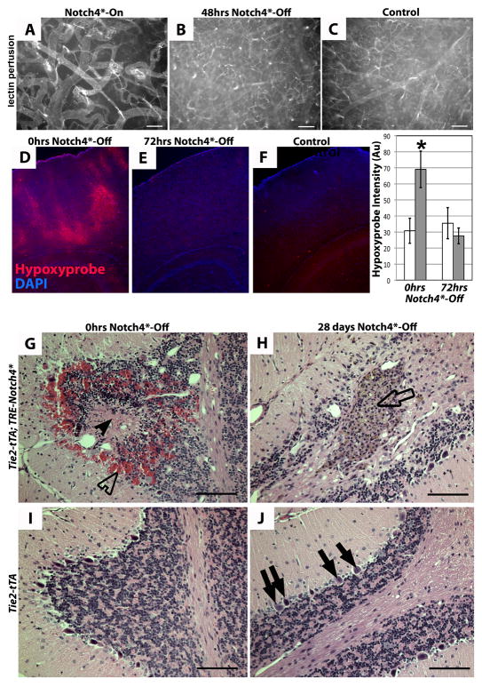
Figure 6. Repression of Notch4* normalizes vascular perfusion, oxygenation, tissue structure.
(A–C) Vascular perfusion of surface vessels of the cerebral cortex by fluorescent tomato-lectin. Following repression of Notch4*, capillary perfusion was increased. (D–F) Immunofluorescence (red) staining for hypoxyprobe (pimonidazole) adduct in coronal section of mouse cortex. Patches of staining were visible in mutant mice with neurologic defects before Notch4* suppression (D). Staining was reduced 72hrs after suppression of Notch4* (E). Control tissue shows an absence of staining (F). Quantification of staining intensity in cortical brain relative to non-specific IgG controls (*P<0.05 vs. all other groups). n=9 at 0hrs Notch4*-Off, n=8 at 24hrs Notch4*-Off. (G–J) Hematoxylin and eosin staining of sagittal paraffin sections of cerebellum. In Notch4*- mutant prior to Notch4* repression (0hrs Notch4*-Off), areas of hemorrhage (open arrowhead) and necrotic tissue (closed arrowhead) were visible (G). After 28 days of Notch4* suppression (Notch4*-Off), areas of scarring were visible (open arrow), but hemorrhage and necrotic tissue had been resolved (H). The numbers of purkinje cells were decreased in H when compared to these cells in the corresponding area in control J (solid arrows). Granular cells (open arrow in H) were found in the scarred area. Scale bar = 100μm.

