Figure 5.
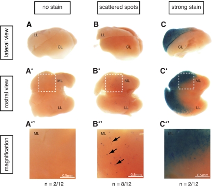
CNC12 activates transcription in the mouse E14.5 liver. Twelve embryos resulting from Hsp68CNC12 LacZ transgenesis showed blue staining in the liver or other structures, indicating successful transgene integration. The livers of these 12 embryos were dissected and pictures from lateral (a-c) and rostral (a'-c') views were taken for each of them (1 representative liver is shown per group; see supplemental Figure 2 for the complete set). As shown in panel a-a″, b-b″, and c-c″, 3 groups could be detected within the 12 transgenics embryos: (a-a″) embryos with no lacZ transcriptional activation in the liver (n = 2), (b-b″) embryos with scattered blue spots specifically in the liver (n = 8), and (c-c″) embryos exhibiting a strong staining in the median and left lobes of the liver (n = 2). Panel a″-c″ are magnifications of the median lobe of each of the representative livers (white boxed region in each of the liver from a'-c' panel). LL: left lobe, CL: caudate lobe, ML: median lobe.

