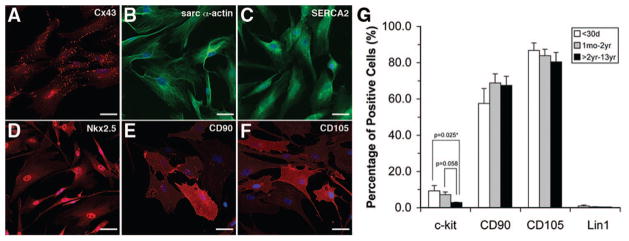
Figure 5.
Confocal microscopy images of CDC at late passage (P4) are shown. A through C, Expression of cardiomyocyte-specific markers. A, Clusters of CDCs expressed connexin 43 (Cx43). The majority of CDC expressed α-sarcomeric actin (α-actin) and SERCA2 (B and C). CDCs at P4 also expressed NKX2–5 (D) and mesenchymal markers CD90 and CD105 (E and F). Flow cytometry analysis of CDC at P4 (G). CDCs were negative for hematopoietic lineage (Lin1) and positive for CD90 (65 ± 5%) and CD105 (81 ± 3%). C-kit+ CDCs were higher in neonates (9 ± 3%) and infants (7 ± 2%) compared with children >2 years of age (3 ± 0.3%). Coexpression of c-kit with CD90 and CD105 was observed (45% to 64%) in the majority of patient samples with no difference between patient age groups. Error bars are ±SEM.

