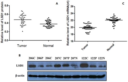
Figure 2. Up-regulation of LSD1 expression in NSCLC tumor tissues.
(A) The protein level of LSD1 in lung cancer tissue was significantly higher than in normal tissue (P<0.01), as detected by Western blot. β-tubulin was used as a loading control. (B) Sample 122, 206 and 207 are shown as representatives of the three groups: N, normal lung tissues: C, NSCLC tissues; P, paracarcinoma tissues. (C) The mRNA level of LSD1 was detected by qRT-PCR, and the mRNA expression of LSD1 was significantly higher in tumor tissue than in normal tissue (P<0.05).

