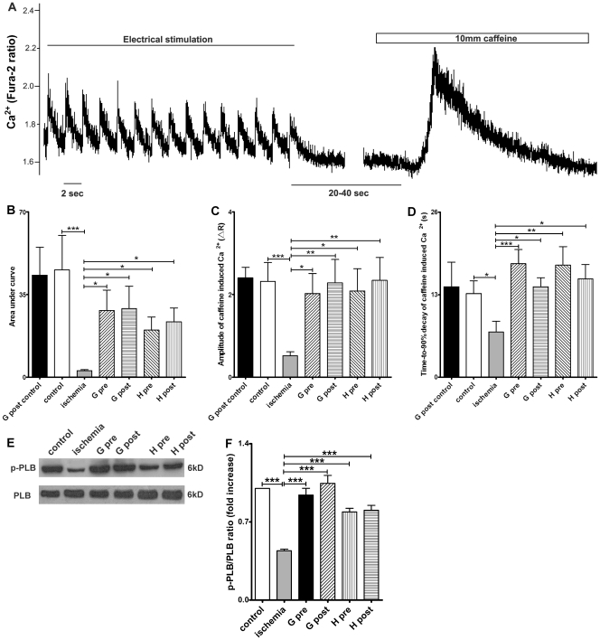
Figure 4. Effects of GHS on sarcoplasmic reticulum (SR) Ca2+ content and phospho-phospholamban (p-PLB)/phospholamban (PLB) expression.
(A) Illustration of the SR Ca2+ content measurement protocol (data from cardiomyocytes exposed to ischemia/reperfusion). R represents the emission fluorescence ratio of fura-2 from excitation at 340 and 380nm. Cardiomyocytes were perfused with Tyrode solution containing 1.5 mM CaCl2 and paced at 0.5 Hz for at least 30 s. 10 mM caffeine was then added to induce SR Ca2+ release. SR Ca2+ content, as determined by both (B) Area under curve and (C) amplitude of the caffeine-induced Ca2+ release, significantly decreased after 20 min ischemia compared with the control group. Ghrelin (G) or hexarelin (H) pre-treatment (pre) and post-treatment (post) significantly increased the SR Ca2+ content after 20 min ischemia (B and C), but introduction of ghrelin into the perfusion system at 40 min and lasting for 10 min (G post control) had no effect on the SR Ca2+ content of the cells isolated from the normal perfused heart. (D) The time-to-90% decay of caffeine-induced increases in [Ca2+]i mainly reflect the Ca2+ clearance ability of the Na+/Ca2+ exchanger (NCX). n = 18, 44, 44, 33, 38, 41 and 38 cells/3 mice in G post control, control, ischemia, G pre, G post, H pre and H post, respectively. (E) Representative western blots of the total phospholamban (PLB) and the phosphorylated PLB (p-PLB) in 6 groups and (F) the densitometric quantification of ratio of p-PLB/PLB (expressed as fold increase relative to control). n = 5 mice in each group. Data are shown as means ± S.E.M. and analyzed by one-way ANOVA with Tukey’s post hoc test. *P < 0.05, **P < 0.01, *** P < 0.001 vs ischemic group.

