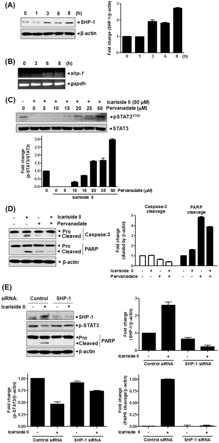
Figure 6. Involvement of protein tyrosine phosphatase SHP-1 in Icariside II-induced apoptosis in U937 cells.
(A) Cells were treated with Icariside II (50 µM) for 0, 1, 3, 6 or 8 h. Cell lysates were prepared and subjected to Western blotting for SHP-1. (B) Total RNA from cells treated with Icariside II (50 µM) 0, 3, 6 or 8 h was extracted, and the mRNA level of SHP-1 was analyzed by RT-PCR. GAPDH was used as an internal control. (C) Cells were treated with Icariside II (50 µM) in the absence or presence of pervanadate (0, 5, 10, 15, 20, 25 or 50 µM) for 9 h. Cell lysates were prepared and subjected to Western blotting for phospho-STAT3 and STAT3. (D) Cells were treated with Icariside II (50 µM) and/or pervanadate (20 µM) for 9 h. Cell lysates were prepared and subjected to Western blotting for caspase-3 and PARP. Representative results of three independent experiments are shown for each experiment. (E) Cells were transiently transfected with either SHP-1 or scrambled siRNA (40 nM) for 48 h and then treated with Icariside II (50 µM) EP for 9 h. Western blotting was performed for SHP-1, phospho-STAT3 and PARP.

