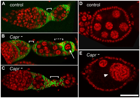
Figure 2. Characterization of egg chamber and stalk defects in Capr- ovaries.
A-C) Germarium, budding egg chamber, and stalk (white bracket) of the indicated genotypes stained with antibodies to the follicle cell marker, FASIII (green), and with TO-PRO-3 iodide (red). A) Df(3L)Cat/+ (Control), B) Capr- showing a reduced primary stalk, and an aberrantly packaging egg chamber displaying an absence of stalk (dashed bracket) and misencapsulation of a single germline cell (arrow), and C) Capr- containing a disorganized stalk. D-E) Egg chambers stained for TO-PRO-3 (red). Optical sectioning revealed 16 nuclei in the control egg chamber (D), but fewer cells in a Capr- egg chamber (E) including nuclei of inappropriate size for this stage (arrowhead). Scale bar is 30 microns.

