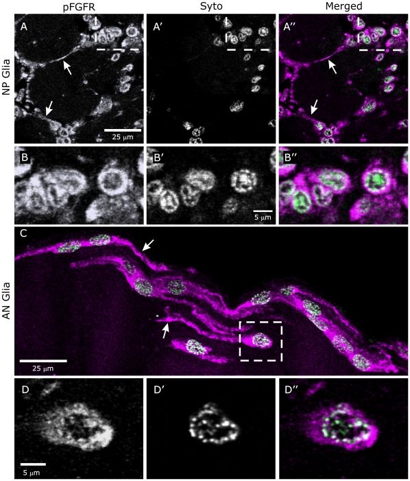
Figure 4. Activated FGFRs are present on glial cell membranes and in nuclei.
A,B: Labeling for pFGFRs is present on NP glial processes (arrows) and associated with glial cell bodies (A, A″). Syto labels glial nuclei. B–B″: An enlarged view of the boxed area of A″ demonstrates co-localization of labels for pFGFRs and DNA in glial cell nuclei. C,D: As for NP glia, AN glia label for pFGFRs both on their processes (arrows in C) and in their nuclei (D″). In both cases colocalization appears to be confined to DNA close to the nuclear envelope (B″, D″). Images are single optical sections (40× objective) except for C (16 µm stack).

