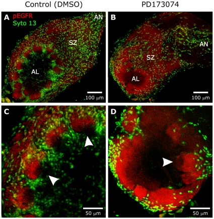
Figure 7. A,B:
Animals treated as in Figure 6A,B and examined at stage 8. Labeling with an antibody to activated Epidermal GFRs (red) reveals a normal pattern of activation in the ORN axons demonstrating that PD173074 did not affect the Epidermal GFRs. C,D: Enlarged images from the same animals demonstrate that the axons occupy their normal territory within the apical half of glomeruli in the control AL (arrowheads in panel 7C) and in the outer portion of the neuropil in the PD173074-treated AL (arrowhead in panel 7D), where glomeruli are demarcated only by glial processes, not by glial cell bodies as in normal lobes. Depth of projections was 10 µm.

