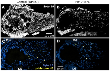
Figure 9. PD173074 treatment results in reduced glial numbers at later stages.
A,B: Syto 59 labeling of the same antennal lobes shown in Figure 8A–D. At lower magnification, Syto 59 labeling of cell nuclei illustrates the significant reduction in cell number in treated animals at later stages (stage 11 shown). The reduction occurs in regions normally occupied solely by glial cells. Blocking glial FGFR activation leads to a decrease in proliferation. C,D: Control and PD173074-treated animals were allowed to develop to late stage 5, then dissected and their brains labeled with an antibody to phospho-histone H3, an indicator of mitosis (yellow). Syto 13 (blue) was used to visualize all glial nuclei. Projection depths = 30 µm in A,B and 10 µm in C,D.

