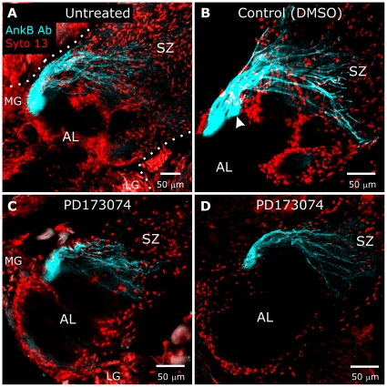
Figure 12. Blocking glial FGFR activation does not greatly perturb targeting of ORN axons to a specified glomerulus.
Brains from untreated animals (A), control (DMSO, B), and animals injected with PD173074 in DMSO beginning at stage 3 (C,D) were labeled at stage 7 with Syto 13 (red) to identify glial nuclei and AL neuron cell bodies. Axons targeting a single glomerulus were labeled with an antibody produced against human Ankyrin B (blue), though the Manduca protein recognized by this antibody is unknown [33]. Although treated animals displayed a lack of NP glial cell body migration in the antennal lobe, axons labeled with the Ankyrin B antibody were seen to project to a single region (C,D) adjacent to the medial group of AL neurons, as they do in untreated and control animals (A,B). Dotted lines in A outline the antennal lobe and AL neuron cell groups (LG & MG). Arrowhead in B points to a large fascicle which merged with the main fascicle to form a single glomerulus in the adjacent section. Projection depths = 30 µm in A, 22.5 µm in B, 12.5 µm in C, 20 µm in D.

