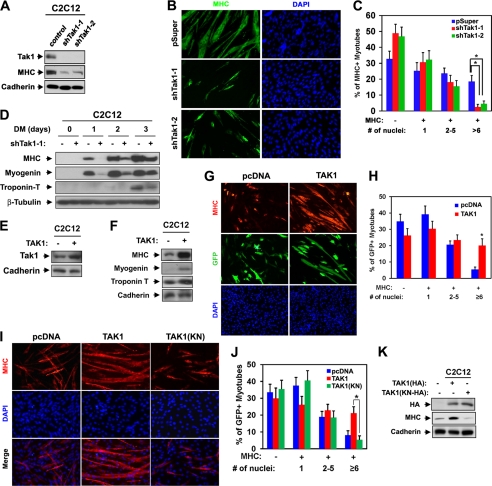
FIGURE 2.
TAK1 promotes myoblast differentiation. A, lysates of C2C12 cells that stably express shTak1-1, shTak1-2, or control (pSuper) vectors at differentiation day 3 (DM3) were immunoblotted with antibodies to TAK1, MHC, and cadherin as a loading control. B, photomicrographs of C2C12 cells shown in A that were cultured in DM for 3 days, fixed, and stained with antibody to MHC. Nuclei were visualized by DAPI staining. C, quantification of myotube formation by the cell lines shown in B. The values represented are the means of triplicate determinations ± 1 S.D. The experiment was repeated three times with similar results. Asterisks indicate difference from the control at p < 0.01. D, lysates of C2C12/shTak1-1 cells from various differentiation times were immunoblotted with antibodies to MHC, myogenin, or troponin T and to β-tubulin as a loading control. E, lysates of C2C12/control or C2C12/TAK1 cells were immunoblotted with antibodies to TAK1 and to cadherin as a loading control. F, lysates of C2C12/control or C2C12/TAK1 cells that were cultured in DM for 2 days were immunoblotted with antibodies to MHC, myogenin, or troponin T and to cadherin as a loading control. G, C2C12 cells were transiently transfected with control or TAK1 expression vector and a GFP expression vector to mark transfectants. Cells at differentiation day 2 were immunostained for MHC (red) and visualized for GFP expression (green). Nuclei were visualized by staining with DAPI (blue). H, quantification of myotube formation shown in G. Values represent the means of triplicate determinations ± 1 S.D. The experiment was repeated three times with similar results. The asterisk indicates difference from the control at p < 0.01. I, C2C12 cells were stably transfected with TAK1, a kinase-negative form of TAK1 (TAK1(KN)), or a control expression vector and differentiated for 2 days followed by immunostaining for MHC (red). Nuclei were visualized by DAPI staining (blue). J, quantification of myotube formation by the cell lines shown in I. Values represent the means of triplicate determinations ± 1 S.D. The experiment was repeated three times with similar results. The asterisk indicates difference from the control at p < 0.01. K, lysates of C2C12 cells that stably express TAK1, TAK1(KN), or control vectors in I were immunoblotted with antibodies to HA and MHC and to cadherin as a loading control.

