FIGURE 5.
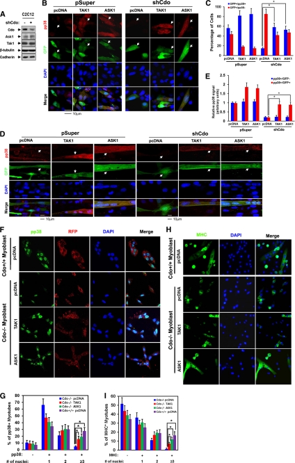
TAK1 and ASK1 rescue defective p38MAPK activation and myotube formation of Cdo-depleted myoblasts and Cdo−/− myoblasts. A, lysates of C2C12/pSuper and C2C12/shCdo cells were immunoblotted with antibodies to Cdo, ASK1, and TAK1 and to β-tubulin and cadherin as loading controls. B, C2C12/pSuper and C2C12/shCdo cells were transiently transfected with ASK1(FLAG), TAK1(HA), or control expression vector plus a GFP vector to mark the transfectants, and 2 days later cells were immunostained with antibodies to pp38 (red) and visualized for GFP expression (green). Cell nuclei were visualized by staining with DAPI (blue). The white arrowheads indicate transfected cells. C, quantification of cultures shown in B. GFP+ cells were scored as positive or negative for pp38 staining. Values represent the means of triplicate determinations ± 1 S.D. The experiment was repeated three times with similar results. Asterisks indicate difference from the control at p < 0.01. D, C2C12/pSuper and C2C12/shCdo cells were transiently transfected with ASK1(FLAG), TAK1(HA), or the control expression vector and a GFP vector to mark the transfectants. Cells at differentiation day 2 were fixed and immunostained with antibodies to pp38 (red) and visualized for GFP expression (green). White arrowheads indicate transfected cells. E, quantification of cultures shown in D. The intensity of the immunofluorescent pp38 signals in GFP+ and GFP− cells was quantified with the values obtained from control vector-transfected cultures set to 1.0. Values represent the means of triplicate determinations ± 1 S.D. The experiment was repeated three times with similar results. Asterisks indicate difference from the control at p < 0.01. F, Cdo−/− and Cdo+/+ myoblasts were transiently transfected with ASK1(FLAG), TAK1(HA), or control expression vector and an RFP vector to mark the transfectants. Cells were induced to differentiate for 1 day by removing bFGF, immunostained with antibodies to pp38 (green), and visualized for RFP expression (red). Cell nuclei were visualized by staining with DAPI (blue). G, quantification of pp38-positive multinucleated myotubes shown in F. Values represent the means of triplicate determinations ± 1 S.D. The experiment was repeated three times with similar results. Asterisks indicate difference from the control at p < 0.01. H, Cdo−/− and Cdo+/+ myoblasts were transfected with expression vectors for ASK1(FLAG), TAK1(HA), or the control. Cells were induced to differentiate for 1 day in DM, fixed, and stained with antibodies to MHC (green). I, quantification of MHC-positive multinucleate myotubes shown in H. Values represent the means of triplicate determinations ± 1 S.D. The experiment was repeated three times with similar results. Asterisks indicate difference from the control at p < 0.01.

