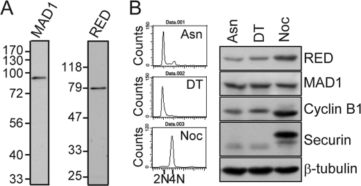
FIGURE 1.
Antibodies against MAD1 and RED (A). Asynchronous HeLa cell lysates were subjected to SDS-PAGE and processed for Western blotting using an affinity-purified rat anti-MAD1 or rabbit anti-RED antibody as a probe. B, protein levels of RED in early S and M phases. Cells were treated with a double thymidine (DT; thymidine, 2 mm) block to arrest them in early S phase or with nocodazole (Noc, 50 ng/ml) to arrest them in M phase. Nocodazole-arrested mitotic cells were shaken off from the Petri dish and collected for Western blot analysis. Asynchronous cell lysates (Asn) were used for comparison. Fifty micrograms of cell lysates were used for Western blotting with antibodies against RED, MAD1, cyclin B1, securin, and β-tubulin (loading control). Cells were also collected, fixed with cold ethanol, and stained with propidium iodide to determine DNA content by flow cytometric analysis.

