FIGURE 1.
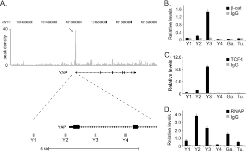
β-Catenin, TCF4, and RNAP bind the first intron of the YAP gene in vivo. A, diagram of the YAP gene locus and the β-catenin-enriched regions identified in a ChIP-Seq screen. Top, gray vertical lines represent peak density plot from the β-catenin ChIP-Seq screen. The arrow indicates the accumulated ChIP-Seq reads that mapped near the YAP transcription start site. Bottom, enlargement of the 5′-end of the YAP1 gene with Y1, Y2, Y3, and Y4 indicating the position of the PCR primer sets used in B, C, and D. B, quantitative real-time PCR analysis of DNA fragments isolated in ChIP assays conducted in HCT116 cells using β-catenin specific antibodies or an irrelevant IgG as a control. The primer sets Y1-Y4 were used to detect β-catenin binding and primers designed against regions in GAPDH (Ga.) and Tubulin (Tu.) were used as negative controls. C and D, as in B except anti-TCF4 antibodies were used in C and anti-RNAP antibodies were used in D to detect TCF4 and RNAP binding in ChIP assays, respectively. In B, C, and D, error is S.E.

