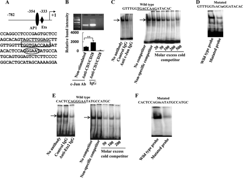
FIGURE 1.
c-Jun and Ets2 bind to the proximal SYK promoter. A, the SYK proximal promoter area is depicted here. The proposed AP-1 and Ets binding sites are marked. B, 3 × 10⁶ normal T cells were cultured in the presence or absence of anti-CD3/CD28 for 30 min and then lysed and used for ChIP experiments with anti-c-Jun or control antibody (Ab) as described under “Experimental Procedures.” The immunoprecipitated DNA was amplified using primers specific for the SYK proximal promoter. A representative experiment and cumulative data from three independent experiments are shown here. C, resting normal T cells were lysed, and the nuclear extract was incubated with a double-stranded oligonucleotide spanning the −354 region of the proximal SYK promoter (−354 probe). In the left panel, we show an EMSA assay of the nuclear protein/−354 probe solution in the absence of antibody or in the presence of control IgG or anti-c-Jun antibodies. In the right panel, the same EMSA nuclear protein/−354 probe solution was incubated in the presence of various concentrations of nonradiolabeled −354 probe, and the autoradiographic picture is shown. D, resting normal T cells were lysed, and the nuclear extract was incubated with the −354 probe or a mutated double-stranded oligonucleotide spanning the −354 region of the proximal SYK promoter (mutated −354 probe); the autoradiographic picture is shown here. E, resting normal T cells were lysed, and the nuclear extract was incubated with a double-stranded oligonucleotide spanning the −333 region of the proximal SYK promoter (−333 probe). In the left panel, we show an EMSA assay of the nuclear protein/−333 probe solution in the absence of antibody or in the presence of control IgG or anti-Ets2 antibodies. In the right panel, the same EMSA nuclear protein/−333 probe solution was incubated in the presence of various concentrations of nonradiolabeled −333 probe, and the autoradiographic picture is shown. F, resting normal T cells were lysed, and the nuclear extract was incubated with the −333 probe or a mutated double-stranded oligonucleotide spanning the −333 region of the proximal SYK promoter (mutated −333 probe); the autoradiographic picture is shown here.

