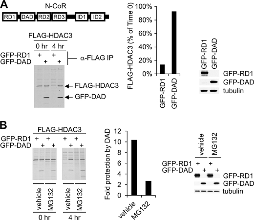
FIGURE 2.
Role of the corepressor DAD domain in the regulation of HDAC3 stability. A, pulse-chase analysis of FLAG-HDAC3 turnover in 293T cells transfected with FLAG-HDAC3 along with GFP-RD1 or GFP-DAD. The middle panel shows the quantification of the remaining HDAC3 at the 4-h time point as a percentage of the initial HDAC3 level at the 0-h time point. The right panel is a Western blot analysis of whole cell extracts showing the expression of GFP-RD1 and GFP-DAD in 293T cells transfected with FLAG-HDAC3 along with GFP-RD1 or GFP-DAD. GFP-RD1 and GFP-DAD were detected using the anti-GFP antibody. B, pulse-chase analysis performed as described in A in the presence of MG132 (20 μm) or dimethyl sulfoxide (DMSO; vehicle control). The middle panel shows the ratio of remaining HDAC3 percentage at 4-h time point in DAD-transfected to RD1-transfected cells in the presence of MG132 or dimethyl sulfoxide. The right panel is a Western blot analysis of cell extracts showing the expression of GFP-RD1 and GFP-DAD in vehicle (dimethyl sulfoxide)-treated and MG132-treated 293T cells transfected with FLAG-HDAC3 along with GFP-RD1 or GFP-DAD. GFP-RD1 and GFP-DAD were detected as described in A. Cells were treated with dimethyl sulfoxide or MG132 for 6 h before lysis.

