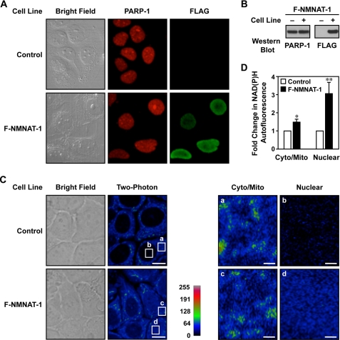
FIGURE 2.
NMNAT-1 localizes to nucleus and mediates nuclear NAD+ synthesis in MCF-7 cells. A, immunofluorescent staining of PARP-1 and NMNAT-1 in control and FLAG-NMNAT-1 (F-NMNAT-1)-expressing MCF-7 cells. B, Western blot analysis of PARP-1 and FLAG-NMNAT-1 protein levels in the MCF-7 cells used in A. C, ectopic expression of NMNAT-1 increases nuclear NAD(P)H autofluorescence in MCF-7 cells. Left, bright field and two-photon excitation microscopy images are shown for control and FLAG-NMNAT-1-expressing MCF-7 cells. Scale bars, 10 μm. Right, detailed images showing the two-photon microscopy signals from the cytoplasm/mitochondria (cyto/mito) and nucleus (nuclear) for control and FLAG-NMNAT-1-expressing MCF-7 cells. Scale bars, 1 μm. The white boxes labeled a–d in the left panels represent the regions labeled a–d magnified in the right panels. D, quantification of NAD(P)H autofluorescence levels from two-photon excitation images. Three to four different fields, each with seven to 10 cells, were analyzed for each condition. The bars shown represent the mean, and the error bars represent S.E. Statistical significance was determined using a two-tailed Student's t test. *, p value <0.1; **, p value <0.05.

