FIGURE 3.
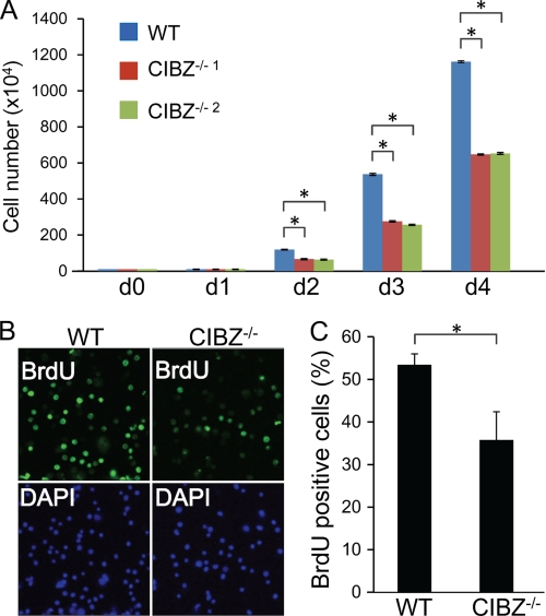
CIBZ deletion in ESCs resulted in reduced proliferation. A, analysis of cell growth in cultures of CIBZ−/− and control ESCs were seeded into 6-cm plate (1 × 10⁵ cells/well). Cell number was counted daily after the seeding (d 0). Results are mean ± S.E. of three independent experiments (n = 6 in total, *, p < 0.05). B and C, knock-out of CIBZ resulted in reduced BrdU incorporation in CIBZ−/−1 ESCs at 3 days after seeding. B, representative fluorescence microscopy images of BrdU- or DAPI-positive cells. C, histogram showing percentages of BrdU- and DAPI-positive cells in each culture. The data shown are representative of 10 randomly chosen fields. Results are mean ± S.E. of three independent experiments. *, p < 0.05.

