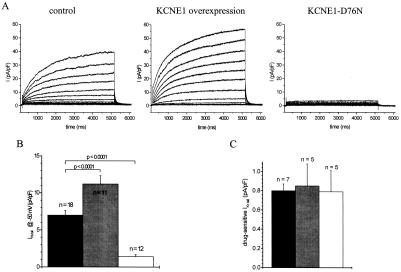
Figure 5.
IKs and IKr current density in control guinea-pig ventriculocytes compared with myocytes that were infected in vivo with AdCGI-KCNE1 or AdCGI-KCNE1-D76N. (A) Original current traces recorded with 5-s depolarizing steps from 60 mV to −20 mV demonstrate that enhanced expression of KCNE1 increased IKs current size, whereas KCNE1-D76N substantially suppressed IKs. (B) Mean IKs tail current density measured at −50 mV after depolarization to 60 mV was increased by KCNE1 overexpression (gray bar) by ≈60% and reduced by KCNE1-D76N (white bar) by ≈80% compared with noninfected cells (black bar). (C) IKr drug-sensitive tail current density was measured as described in Fig. 1. We did not observe any effect of KCNE1 overexpression (gray bars) or KCNE1-D76N (white bars) compared with noninfected myocytes (black bars) on drug-sensitive IKr tail current density in guinea-pig myocardium.

