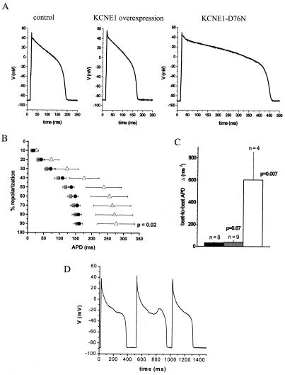
Figure 6.
Effect of in vivo expression of KCNE1 wild-type and KCNE1-D76N on cardiac repolarization in freshly isolated guinea pig myocytes. IKs increase by KCNE1 overexpression (gray) exhibited no significant effect on overall APD (APD90 154.3 ± 10.4 ms, n = 9; P = 0.49) (A and B) or beat-to beat APD variability (C) compared with noninfected myocytes (black). However, IKs suppression by KCNE1-D76N (white) prolonged overall APD almost 2-fold (APD90 277.1 ± 58.7 ms, n = 7; P = 0.02) (A and B) and resulted in an extensive increase in beat-to-beat APD variability (C), which led to frequent arrhythmogenic EADs (D).

